
Scientists who cut the brain of a deceased 65-year-old woman into 7,400 slices have created a 3D digital atlas of the human brain whose resolution is 50 times greater than that of previously existing models.
Called “BigBrain,” the project was published in the latest issue of the US journal Science.
The model shows features finer than a human hair and almost on the scale of individual cells, said lead researcher Katrin Amunts, a neuroscientist at Germany’s Juelich Research Centre and a professor at the University of Dusseldorf. Those two institutions, along with the Montreal Neurological Institute and Hospital, provided the bulk of the research team.
“Although the cells are still somewhat blurred, we see how densely packed they lie and how they’re distributed. We see into the furthest corner of the brain,” Amunts said.
She made an analogy by way of illustration. While continents, countries and cities were recognizable in old brain atlases, “now we can look into individual streets.” She said the brain model could later be supplemented with data on molecular structure, genetic information or connections between brain regions.
The scientists found that the cells were arranged according to brain functions. “The arrangement depends on whether the region controls movement, sounds or light signals,” Amunts said.
The BigBrain atlas of brain cell architecture aims to facilitate important insights into processes such as cognition, language and emotions. Scientists also want to understand why these processes sometimes go wrong.
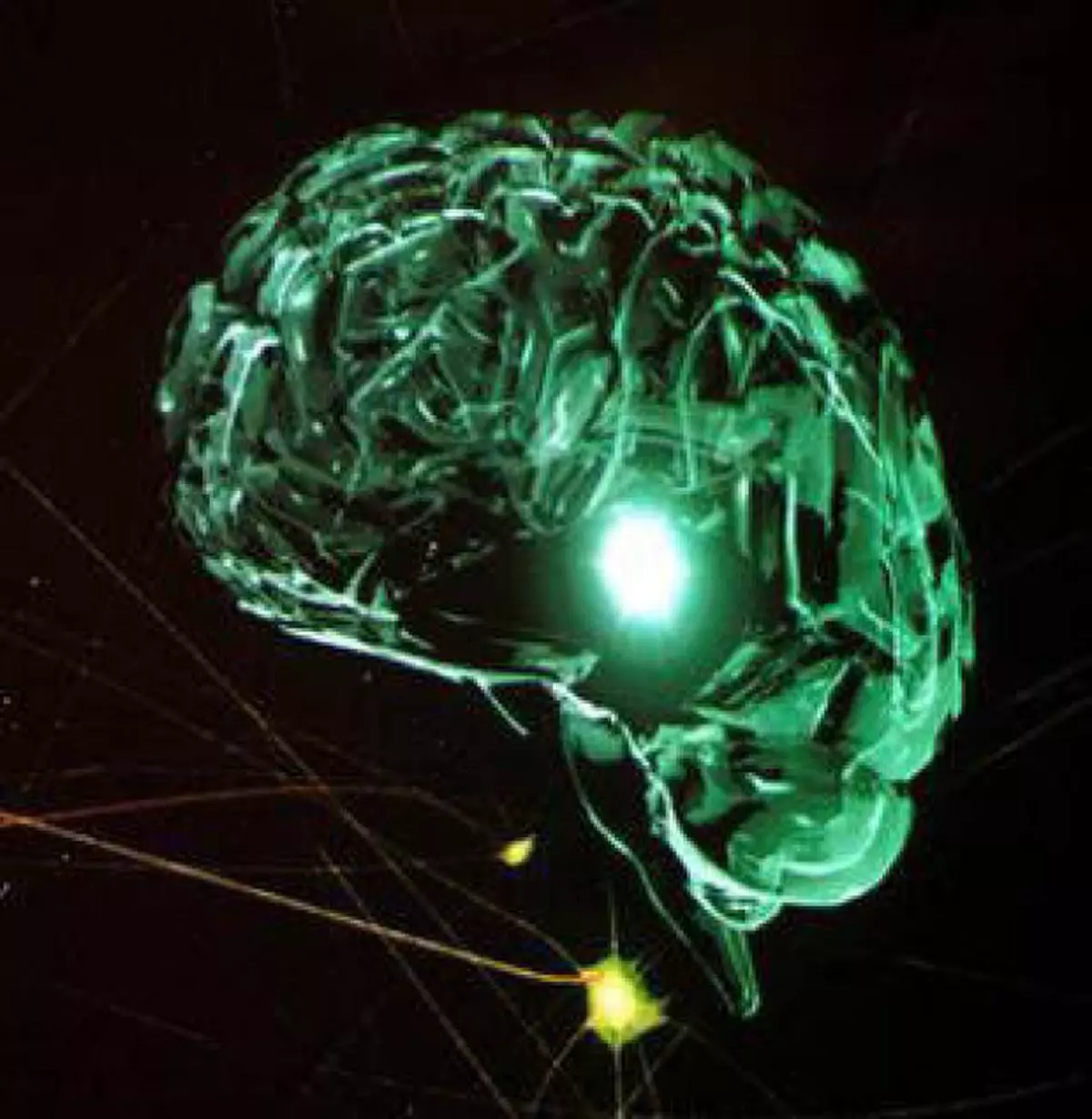
BigBrain will help doctors too, Amunts said. During deep brain stimulation of patients with Parkinson’s Disease, for example, exact placement of the merely two-millimetre-thick electrodes is important, she remarked.
“The atlases that are used for this are very imprecise in parts,” she said, adding that BigBrain could also be used in cases of other neurological disorders.
According to Amunts, very few laboratories in the world are capable of slicing an entire brain in ultra-thin pieces of consistent quality, but Germany has a long tradition of scientifically preparing the brains of deceased persons.
Another challenge was to develop software to correct small errors in the digitized images due to tears or folds in the extremely thin sections of brain tissue.
![]() Comments
Comments

Comments
Comments have to be in English, and in full sentences. They cannot be abusive or personal. Please abide by our community guidelines for posting your comments.
We have migrated to a new commenting platform. If you are already a registered user of TheHindu Businessline and logged in, you may continue to engage with our articles. If you do not have an account please register and login to post comments. Users can access their older comments by logging into their accounts on Vuukle.